Syoji Kobashi
Professor | Ph.D. in Engineering
Artificial Intelligence and Informatics Course
Data Science Research Group
Professor Kobashi's teaching spans programming, data science, and medical engineering, with an emphasis on cultivating independent thinking and a proactive approach to problem-solving. His research lies at the intersection of artificial intelligence and medicine, developing computational methods that support clinical diagnosis and treatment planning and help anticipate disease onset and prognosis, with the goal of enabling more personalized and effective patient care.
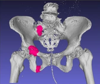
Fracture Detection from CT Images
What students can learn
Students gain hands-on experience in AI-based image analysis, with a focus on computer vision techniques for object detection, while building the interdisciplinary communication skills needed to collaborate effectively with clinical partners.
This project develops AI algorithms to help identify pelvic fractures in CT scans. In older adults, osteoporosis-related pelvic fractures often produce few noticeable symptoms and can go unnoticed without prompt access to a specialist. By supporting fracture assessment in settings such as regional clinics, night-time emergency care, and areas with limited specialist availability, this research aims to help reduce missed diagnoses and enable timely treatment.
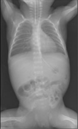
Predicting Chronic Lung Disease in Newborns
What students can learn
Through this project, students develop skills in image-based classification and regression using computer vision, while learning to communicate effectively across disciplines in a medical research setting. The work also serves as a concrete case study in modeling disease onset from clinical imaging data.
Premature infants in neonatal intensive care units often have underdeveloped lungs and face an elevated risk of chronic lung disease. This research explores the use of chest X-ray images taken shortly after birth to predict, at 36 weeks, which infants are at high risk, with the aim of supporting earlier intervention and helping to reduce disease severity or, in some cases, prevent its onset.